Animal Cell Free printable to label +
To draw a well labelled diagram of an animal cell, the cell membrane has to be drawn. The cell membrane is an integral part of the cell structure that keeps the entire cell bound together. It is a phospholipid bilayer surrounding the entire cell with cytoplasm and the organelles in the cell cytoplasm. The membrane is selectively permeable only.

Biology Animal Cell Model Labeled / Image Of An Animal Cell Diagram With Each Organelle Labeled
Animal Cell Diagram & Anatomy Biology Science Animal Cell Label Me! Printout Plant Cell Anatomy Animal Cell Anatomy The cell is the basic unit of life. All organisms are made up of cells (or in some cases, a single cell). Most cells are very small; in fact, most are invisible without using a microscope.

What is a cell? Facts
Diagram of an animal cell Doc Sonic Structures Unique to Animal Cells While animal cells do not have a cell wall, chloroplasts, or a large vacuole, they do have one component plant cells do not. Centrioles: Animal cells contain organelles known as centrioles, which are not present in plant cells.
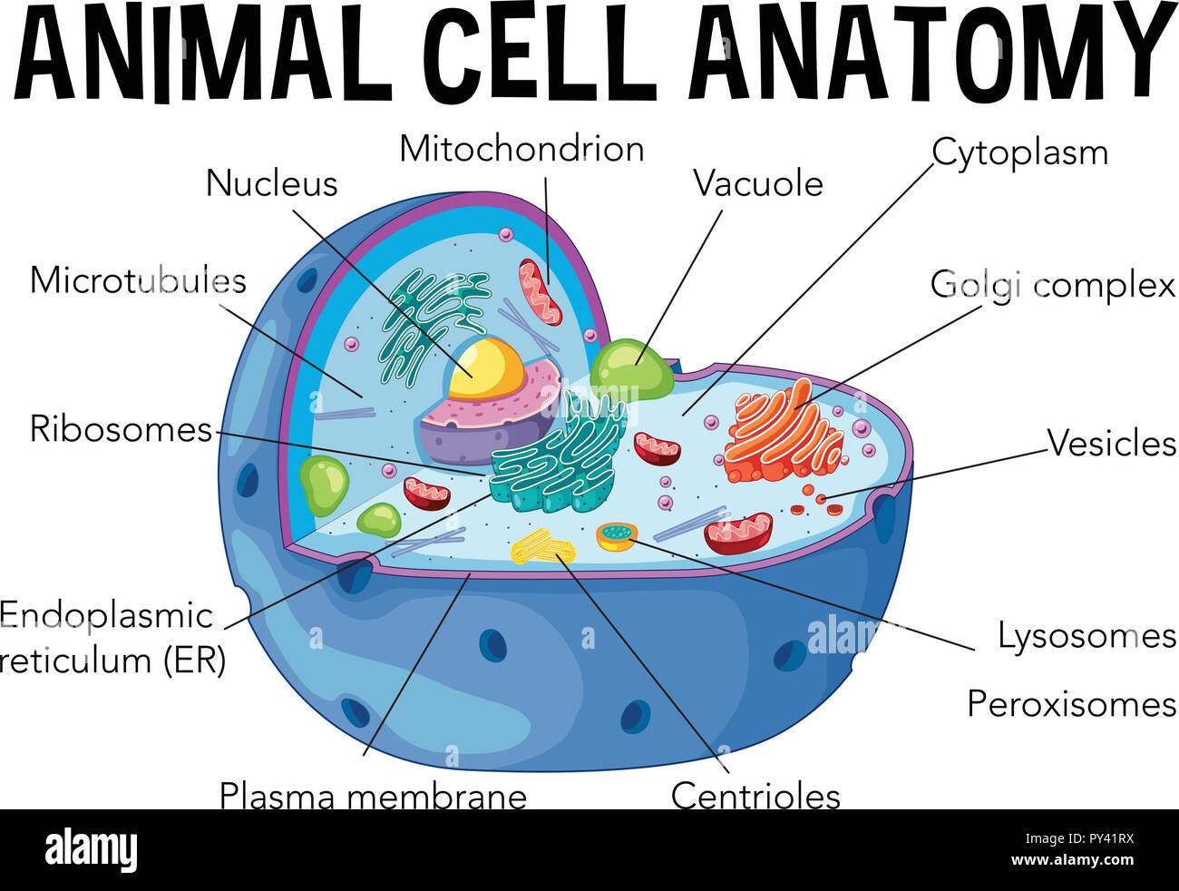
Diagram of animal cell anatomy illustration Stock Vector Image & Art Alamy
Diagram of Animal Cell Read A diagram of an animal cell is useful for understanding the structure and functioning of an animal. This article includes a well-labeled diagram and a brief description of each component of an animal cell. Animal cells are eukaryotic cells with a membrane-bound nucleus.

Cell Structure Animal cell, Animal cell structure, Animal cells worksheet
There are six animal cell diagrams to choose from. The first is a colored and labeled cell diagram. The next is a black and white version of the first. These printables a free for subscribing members of Tim's Printables. Already a member? Please remember to log in. Not yet a member? Join today!

Pictures Of The Animal Cells ANIMALSD
A Labeled Diagram of the Animal Cell and its Organelles There are two types of cells - Prokaryotic and Eucaryotic. Eukaryotic cells are larger, more complex, and have evolved more recently than prokaryotes. Where, prokaryotes are just bacteria and archaea, eukaryotes are literally everything else.
Discovery and Structure of Cells Biology Visionlearning
Definition Animal cells are the basic unit of life in organisms of the kingdom Animalia. They are eukaryotic cells, meaning that they have a true nucleus and specialized structures called organelles that carry out different functions.

Biology 2e, The Cell, Cell Structure, Eukaryotic Cells OpenEd CUNY
Parts of Animal cell diagram . The Cell Organelles are membrane-bound and present within the cells. There are various organelles present within the cell and are classified into three categories based on the presence or absence of a membrane. Listed below are the Cell Organelles of an animal cell along with their functions.

The Structure and Functions of an Animal Cell
Animal Cell: Structure, Parts, Functions, Labeled Diagram June 6, 2023 by Faith Mokobi Edited By: Sagar Aryal An animal cell is a eukaryotic cell that lacks a cell wall, and it is enclosed by the plasma membrane. The cell organelles are enclosed by the plasma membrane including the cell nucleus.
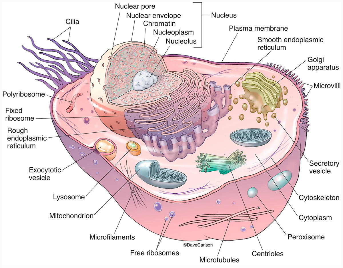
Animal Cell Structure Carlson Stock Art
Learn about the structure and function of animal cells, the basic unit of life in animals. Explore the various organelles and their roles in maintaining homeostasis.. Labeled Diagram. By Go Life Science Posted on December 20, 2022 October 17, 2023. An animal cell is a type of cell that is characteristic of animals and is present in all.
Animal Cell Diagram CBSE Class Notes Online Classnotes123
1. Draw a simple circle or oval for the cell membrane. The cell membrane of an animal cell is not a perfect circle. You can make the circle misshapen or oblong. The important part is that it does not have any sharp edges. [1] Also know that the membrane is not a rigid cell wall like in plant cells.

What Is An Animal Cell? Facts, Pictures & Info For Kids & Students.
Diagram Of Animal Cell Animal cells are eukaryotic cells that contain a membrane-bound nucleus. They are different from plant cells in that they do contain cell walls and chloroplast. The animal cell diagram is widely asked in Class 10 and 12 examinations and is beneficial to understand the structure and functions of an animal.
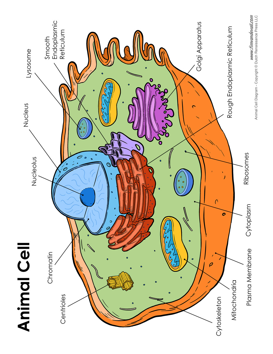
Animal Cell Diagram Labeled Tim van de Vall
Community Structure 17m. 52. Ecosystems 28m. Ecosystems 28m. 53. Conservation Biology 24m. Conservation Biology 24m. Label the structures in this diagram of an animal cell. Review the functions of each of these organelles.

Animal Cell diagram with labels by Russell Kightley Media
The shape of a typical animal cell varies widely from being flat, oval to rod-shaped, while others assume shapes such as curved, spherical, concave, and rectangular. An animal cell ranges in size from 10 to 30 µm. Under the microscope, an animal cell shows many different parts called organelles, that work together to keep the cell functional.

South Pontotoc Biology Plant and Animal Cell Diagrams
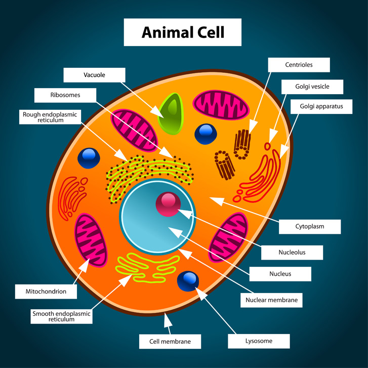
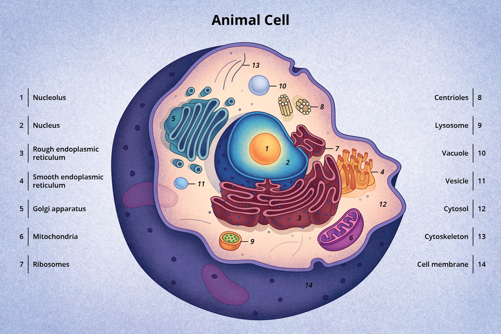
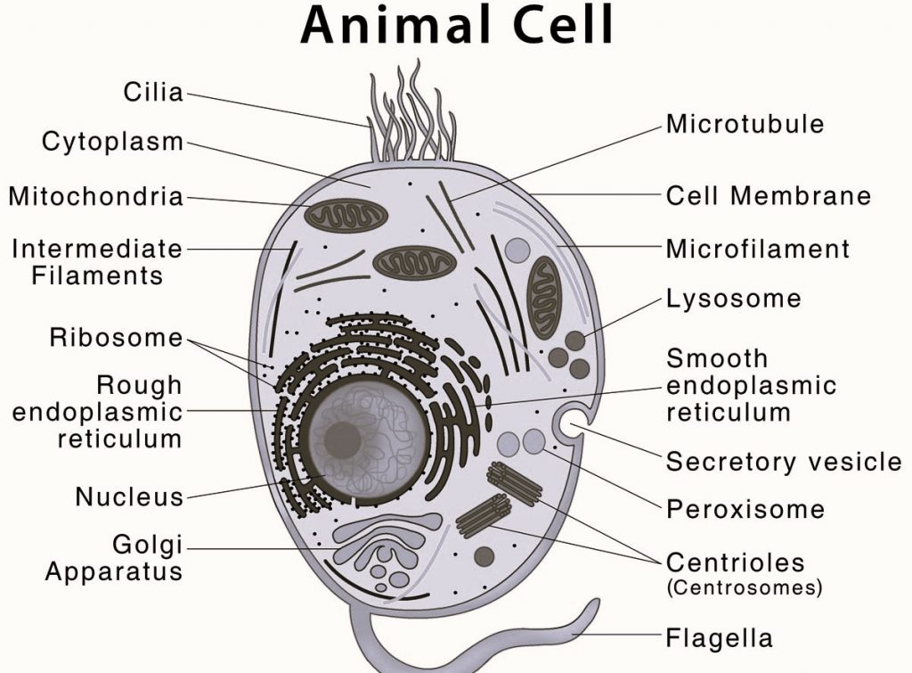
1. Plasma membrane (Cell membrane) 2. Nucleus 3. Cytoplasm 4. Mitochondria 5. Ribosomes 6. Endoplasmic Reticulum (ER) 7. Golgi apparatus (Golgi bodies/Golgi complex) 8. Lysosomes 9. Cytoskeleton Functions of Cytoskeleton 10. Microtubules 11. Centrioles 12. Peroxisomes 13. Cilia and Flagella

Biology 101 Cells Owlcation
Looking for a Labeled Diagram of an Animal Cell? You Found It! What's inside an animal cell? Animal Cells are made up of a number of unique parts, including: Cell Membrane Nucleus Nuclear Membrane Centrosome Lysosome Cytoplasm Golgi Apparatus And more! Create Your Own Animal Cell Diagram - Printable Animal Cell Worksheets and More!